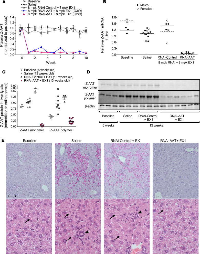
Figure 1. RNAi-mediated reduction of Z-AAT protein and mRNA in young PiZ mouse livers prevented polymer accumulation.
Five-week-old male and female PiZ mice were given IV injections of 8 mg/kg ARC-AAT API plus 8 mg/kg EX1 (RNAi-AAT + EX1) once every 2 weeks (Q2W, n = 24) or once every 3 weeks (Q3W, n = 8) or were given Q2W injections of RNAi-Control plus 8 mg/kg EX1 (RNAi-Control + EX1) (n = 6). Age-matched control animals were injected Q2W with saline (n = 13). Untreated baseline mice were euthanized at 5 weeks of age (n = 5). (A) Plasma was collected at the indicated times and Z-AAT protein measured, shown as the group mean ± SEM relative to pretreatment expression. (B) RNAi-AAT + EX1, RNAi-Control + EX1, and saline-injected mice were euthanized 2 weeks after final injection, and amounts of Z-AAT mRNA in homogenized liver tissue were quantified relative to the geometric mean of the saline group. Males were given 4 injections and euthanized when 13 weeks old. Females were given 5 injections and euthanized when 15 weeks old. (C and D) The amounts of soluble (monomeric) and insoluble (polymeric) Z-AAT protein in liver lysates of male mice at baseline (5 weeks old) or given 4 Q2W injections and euthanized when they were 13 weeks old were measured by semiquantitative Western blotting, shown relative to the saline group as the mean ± SEM, n = 3–10. (E) Representative PAS-D–stained (upper row) and H&E-stained (lower row) liver sections from male mice at baseline or injected Q2W with saline, RNAi-Control + EX1, or RNAi-AAT + EX1. Scale bars for PAS-D indicate 50 μm and for H&E indicate 20 μm. Arrows point to Z-AAT globules and arrowheads to foci of inflammatory cells.

