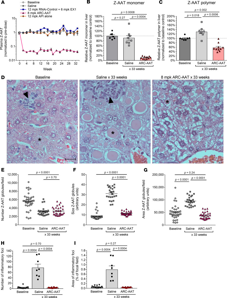
Figure 2. Sustained RNAi-mediated reduction of Z-AAT prevented liver disease and reversed polymer accumulation in adult PiZ mice.
Male PiZ mice, 11–17 weeks old, were given IV injections Q2W of 8 mg/kg ARC-AAT (8 mg/kg ARC-AAT API + 4 mg/kg EX1), 12 mg/kg RNAi-Control plus 6 mg/kg EX1, 12 mg/kg ARC-AAT API alone, or saline (n = 4–12) for 30–31 weeks and were euthanized for evaluation 2 weeks after the final injection. Untreated baseline mice were euthanized at the start of the study. (A) Plasma was collected at the indicated times and Z-AAT protein measured, shown as the group mean relative to pretreatment. (B and C) The amounts of monomeric and polymeric Z-AAT protein in liver lysates were measured by semiquantitative Western blotting, shown relative to baseline for individual animals and as the group means ± SEM. (D) Representative H&E liver sections from PiZ mice at baseline or following 33 weeks of saline (age-matched control) or ARC-AAT treatment. Asterisks indicate globules; arrowheads point to foci of inflammatory cells. Scale bar: 20 µm. (E–G) Z-AAT globules in PAS-D–stained liver sections were quantified in 3 fields of view for each animal (n = 8–12) for the number of globules/field of view (E), the size of globules (F), and the area of the specimens containing globules (G). (H and I) The number of inflammatory foci and area of liver specimens containing inflammatory foci are compared for baseline, saline-injected, and ARC-AAT–treated mice. Means are shown with SEM. Comparisons between groups (B, C, and E–I) were performed using nonparametric Wilcoxon’s rank sum test.

