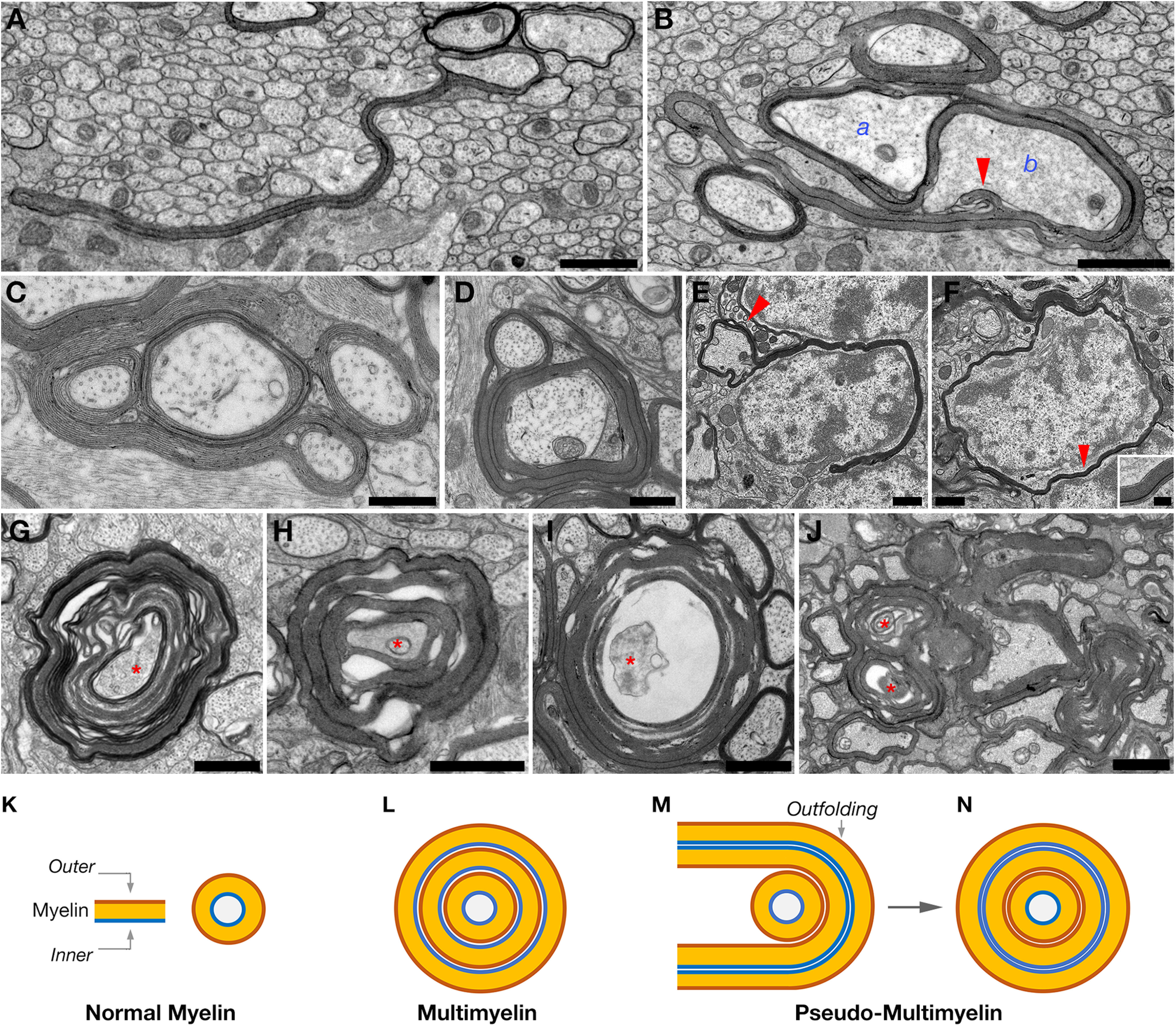
Figure 3.

Cnp-Cre/N-Waspflx/flx mice exhibit severe myelin abnormalities. A, B, EM images of Cnp-Cre/N-Waspflx/flx corpus callosum at P15. The long extending myelin outfolds originated from one axon (labeled a in B) surrounds a nonmyelinated axon (b), and extends an additional outfold (arrowhead) that contact this axon. C, D, High-magnification EM images of optic nerve cross sections obtained from 1-month-old Cnp-Cre/N-Waspflx/flx mutant mice, showing long myelin outfoldings. Myelin outfolds cover other myelinated axons. E, F, Presence of oligodendrocyte myelin around neuronal cell bodies. EM images of the cerebellum of 4-month-old Cnp-Cre/N-Waspflx/flx mutant mice. The presence of myelin outfolds that either partially (E) or completely (F) surrounding neuronal cell bodies. E, Arrow indicates the presence of a nearby myelinated axon. High magnification of area indicated by arrowhead in F showing multiple myelin layers around the cell body. G-I, High-magnification EM images of optic nerve cross sections obtained from P10 (G), P15 (H), and P30 (I) Cnp-Cre/N-Waspflx/flx mutant mice showing different configurations of multimyelinated axons undergoing degeneration and axon (red asterisk) detachment. J, EM image of optic nerve cross sections obtained from 4-month-old Cnp-Cre/N-Waspflx/flx mutant mice showing multiple profiles of focal hypermyelination and degeneration (red asterisk). K–N, Schematic model showing normal myelin (K), multimyelin configuration resulting from aberrant adhesion (L), and pseudo-multimyelin configuration formed by wrapping of a myelin outfolding around myelinated axon (M,N). K, The myelin sheath (Myelin) and the location of the most inner and outer myelin membranes. Scale bars: A, B, D–I, 1 µm; C, 0.5 µm; J, 2 µm.
