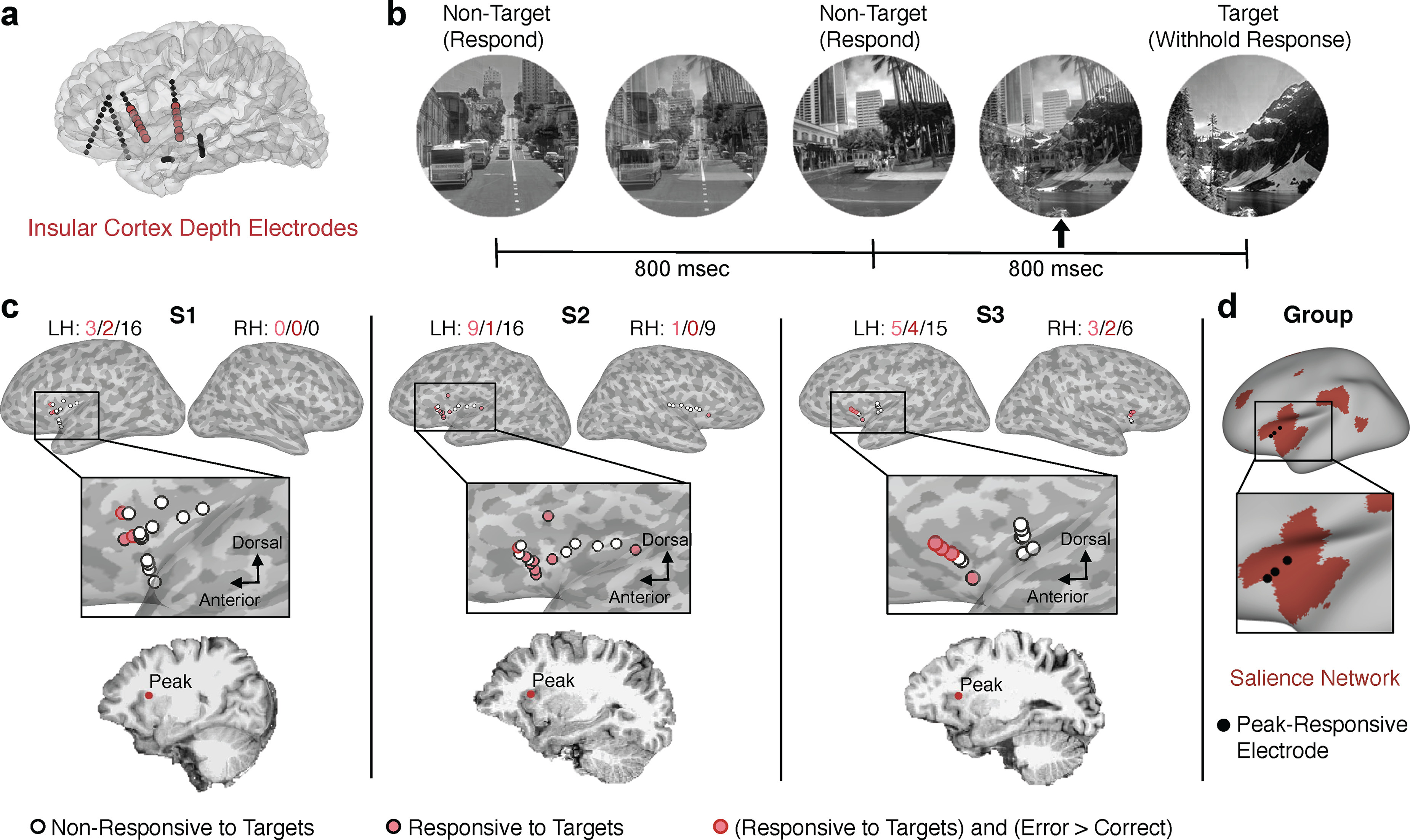
Figure 1.

Task-evoked insula and pupil responses to target stimuli in the GradCPT. a, Depth probes with electrodes implanted in the insula (red) are illustrated in an example subject. b, The GradCPT paradigm. c, Locations of insula electrodes in 3 subjects are shown on the inflated cortical surface. Electrodes were classified into those showing no significant HFB response to GradCPT target relative to nontarget stimuli (white fill, black outline), a significant HFB increased response to target relative to nontarget stimuli (pink fill), and a significant HFB increased response for target trials with correct compared with incorrect behavioral responses (red outline) (Monte Carlo p < 0.05, cluster-based permutation test corrected for number of insula electrodes within subject). The location of the electrode with strongest HFB response within each subject is shown on a 2D sagittal slice. d, Locations of peak-responsive insula electrodes within each subject displayed on the inflated fsaverage cortical surface. Red represents the salience network, based on the fMRI-based Yeo atlas of seven cortical networks.
