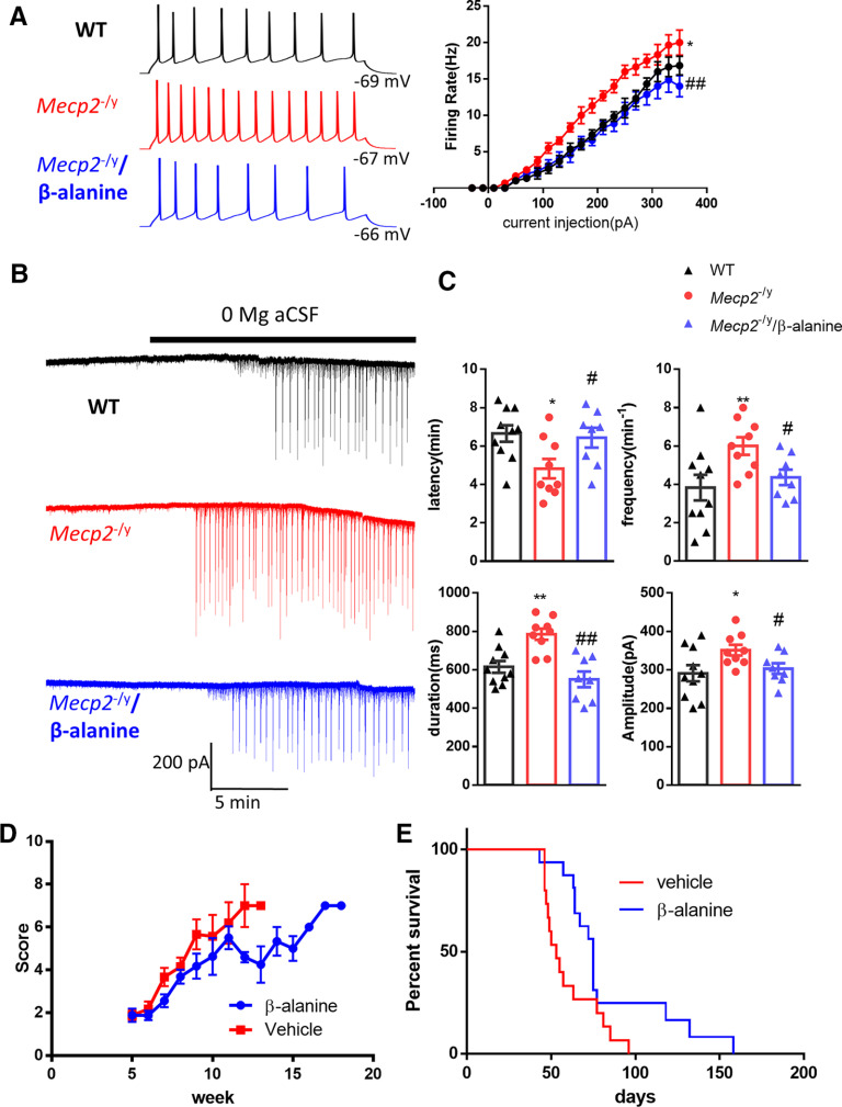
Figure 7.
Inhibition of GAT3 rescued hyperexcitability phenotypes in acute hippocampal slices from the Mecp2 KO mice and alleviated symptoms in Mecp2 KO mice. A, Left, Representative traces showing current injection-evoked action potentials recorded from hippocampal pyramidal neurons in WT (black), Mecp2 KO (Mecp2−/y, red), and Mecp2 KO in the presence of β-alanine (blue). Right, Quantification of the firing rate of hippocampal neurons in response to different current steps. *p < 0.05 versus WT. ##p < 0.01 versus Mecp2−/y. B, Representative traces showing epileptiform activity induced by 0 Mg aCSF in hippocampal slices from WT (top) and Mecp2 KO (Mecp2−/y) mice in the absence (middle) or presence of β-alanine (bottom). C, Quantification of the latency, frequency, duration, and amplitude of the epileptiform activity. The latency is defined as the time elapsed between the application of 0 Mg aCSF and the onset of epileptiform activity. *p < 0.05, **p < 0.01 versus WT. #p < 0.05 versus Mecp2 KO. D, The phenotypic severity scores of male Mecp2 KO (Mecp2−/y) mice receiving daily injection of vehicle or β-alanine. E, Survival curves of male Mecp2 KO (Mecp2−/y) mice receiving daily injection of vehicle or β-alanine.

