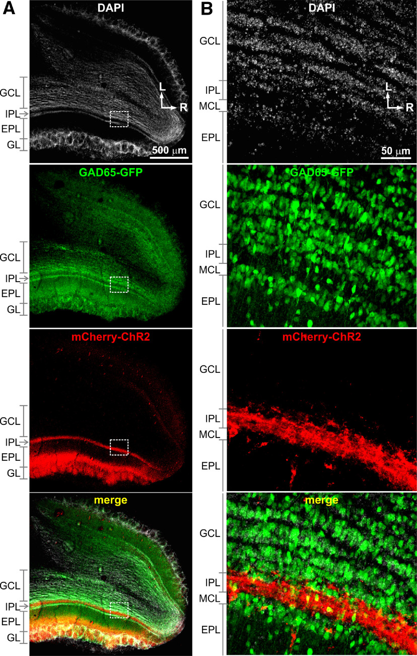
Figure 3.
ChR2-mCherry expression in TCs and GAD65-GFP expression in GABAergic interneurons in the OB. A, The horizontal OB sections prepared from a CCK-Cre x GAD65-GFP mouse with microinjection of AAV2.5-ChR2-mCherry into the medial side of the OB showing DAPI-staining cellular nuclei (white), expression of GAD65-GFP (green) in interneurons including GCs, expression of ChR2-mCherry (red) predominantly in the superficial EPL, GL, and the IPL, which respectively correspond to the locations of STC somata, apical dendrite tufts, and axons, and merged image. B, Blown-up images of the areas labeled in A highlighting expression of GAD65-GFP in GCs in the GC layer (GCL) and ChR2-mCherry expression in cross-sectioned TC axons the IPL. L: lateral; R: rostral.

