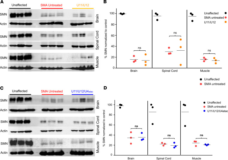
Figure 4. Minor snRNA gene delivery does not increase SMN protein expression in SMA mice.
Tissues for Western blot analysis were collected from brain, spinal cord, and gastrocnemius muscle. Western blot images showing SMN expression levels in tissues collected at P9 from heterozygous (unaffected) control mice as well as untreated and U11/U12- (A) or U11/U12/U4atac-treated (C) SMA mice. No significant changes in the SMN levels were observed in all tested tissues comparing samples from untreated and U11/U12- (B) or U11/U12/U4atac-treated (D) SMA mice. All SMA samples were predictably lower than unaffected samples. Actin was used as a loading control. Comparisons were analyzed by Student’s t test. *P < 0.05. Data expressed as mean ± SEM. n = 3 animals per group. Data in the scatter plots are presented as percentage of SMN normalized to control.

