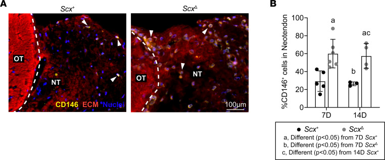
Figure 3. Effect of scleraxis deletion on pericyte density.
(A) Representative immunofluorescence histology demonstrating the presence of CD146+ pericytes in tendons of Scx+ and ScxΔ mice 14D after synergist ablation/plantaris growth procedure. The original tendon (OT) and neotendon (NT) are indicated by the hashed line. CD146, yellow; extracellular matrix (ECM), red; nuclei, blue. Scale bar: 100 μm. White arrowheads indicate the presence of CD146+ pericytes. (B) Quantification of the CD146+ pericytes as a percentage of total cells in the neotendon in Scx+ and ScxΔ mice at 7D or 14D. Values are mean ± SD. Differences between groups were tested using a 2-way ANOVA; a, significantly different (P < 0.05) from 7D Scx+; b, significantly different (P < 0.05) from 7D ScxΔ; c, significantly different (P < 0.05) from 14D Scx+. N ≥ 3 mice per group.

