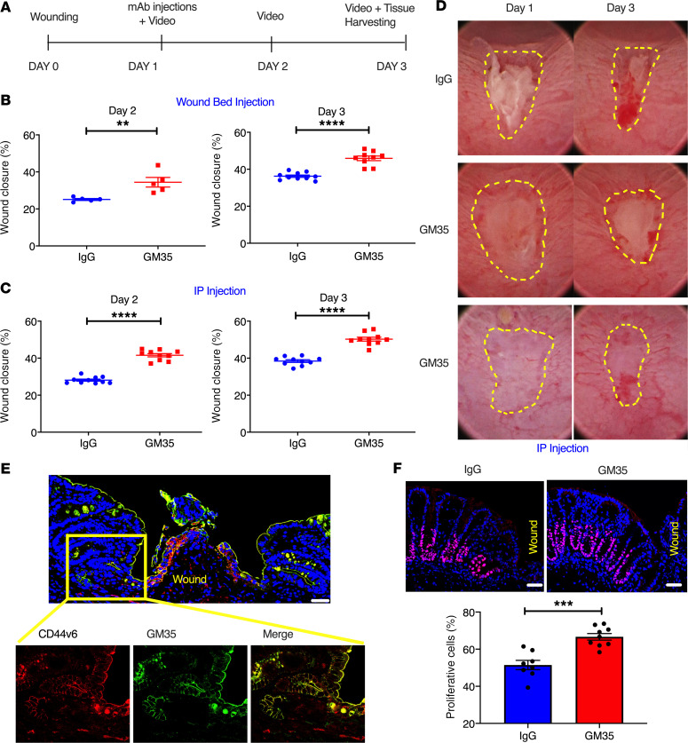
Figure 3. GM35 improves healing of biopsy-induced colonic wounds in vivo.
(A) Schematic overview of experimental design timeline. Animals were wounded on day 0 and treated with GM35 or a control IgG on day 1 via i.p. injection (250 μg/animal) or microinjection directly into the wound bed (10 μg/wound). (B) Percentage of wound healing in animals injected with IgG (blue) or GM35 (red) directly into wound beds. Data are shown as means ± SEM; n = 3 independent experiments. **P < 0.01, ****P < 0.0001. Mann-Whitney U test for day 2 and unpaired t test for day 3. (C) Percentage of wound healing in animals i.p. injected with 250 μg control IgG (blue) or GM35 (red). Data are shown as means ± SEM (n = 3 independent experiments) and were analyzed by 1-way ANOVA followed by Tukey’s post hoc testing. ****P < 0.0001. (D) Representative images of wounds at day 1 and day 3 after i.p. injection with IgG control Ab or GM35. (E) Representative immunofluorescence images of colonic wounds stained with anti-CD44v6 mAb (red) and GM35 (green). Scale bar: 10 μm. Magnification of area adjacent to wound (yellow square) demonstrates expression and colocalization of CD44v6 (red) and sialyl glycan epitopes (GM35, green). (F) Representative immunofluorescence images with proliferation marker Ki67 (pink) in wound-adjacent epithelial crypts after 24 hours of treatment with IgG or GM35. Nuclei are stained in blue. Graph shows quantification of Ki67+ cells in wound-adjacent crypts after treatment with IgG or GM35. Scale bar: 5 μm. Data are shown as mean ± SEM (n = 3 experiments) and were analyzed by 1-way ANOVA followed by Tukey’s post hoc testing. ***P < 0.001.

