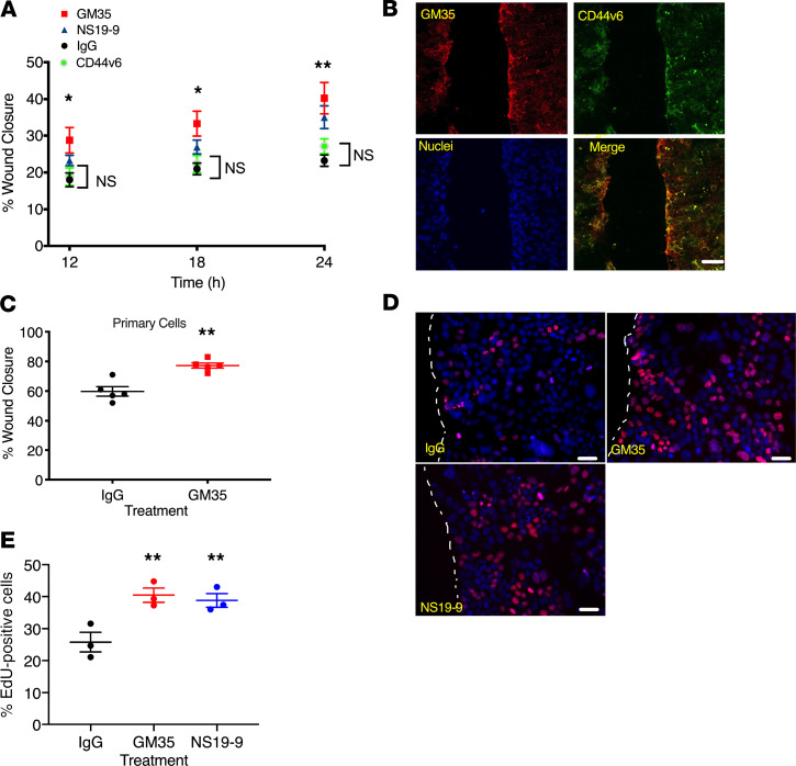
Figure 4. Targeting of sialyl Lewis glycans on CD44v6 with GM35 increases migration and proliferation of human epithelial cells.
(A) Percentage of intestinal epithelial wound closure was calculated by measuring wound widths at 12, 18, and 24 hours after wounding. IECs were incubated with 10 μg/mL GM35, NS19-9, anti-CD44v6 mAb, or IgG matched control antibody. Data are shown as mean ± SEM and were analyzed by 1-way ANOVA followed by Tukey’s post hoc testing (n = 4 experiments, 6 wounds per treatment), *P < 0.05, **P < 0.01. (B) Immunofluorescence staining of scratch-wounded T84 IECs with mAb GM35 in green and anti-CD44v6 mAb in red. Scale bar: 20 μm. (C) Wound closure after 24 hours was measured in colonoid-derived primary human epithelial cells after treatment with 10 μg/mL GM35 or an IgG matched control mAb. Data are shown as mean ± SEM analyzed by 1-way ANOVA followed by Tukey’s post hoc testing (black and red circles represent averages of n = 5 experiments, 6 wounds per group per experiment), **P < 0.01. (D) Scratch-wounded intestinal epithelial monolayers were treated with 10 μg/mL GM35, NS19-9, or control IgG before EdU incorporation was measured 18 hours after injury. Scale bar: 5 μm. (E) Quantification of proliferation/EdU incorporation. Data are shown as mean ± SEM analyzed by 1-way ANOVA followed by Tukey’s post hoc testing (circles represent averages of n = 3 experiments, 5 wounds per treatment); **P < 0.01.

