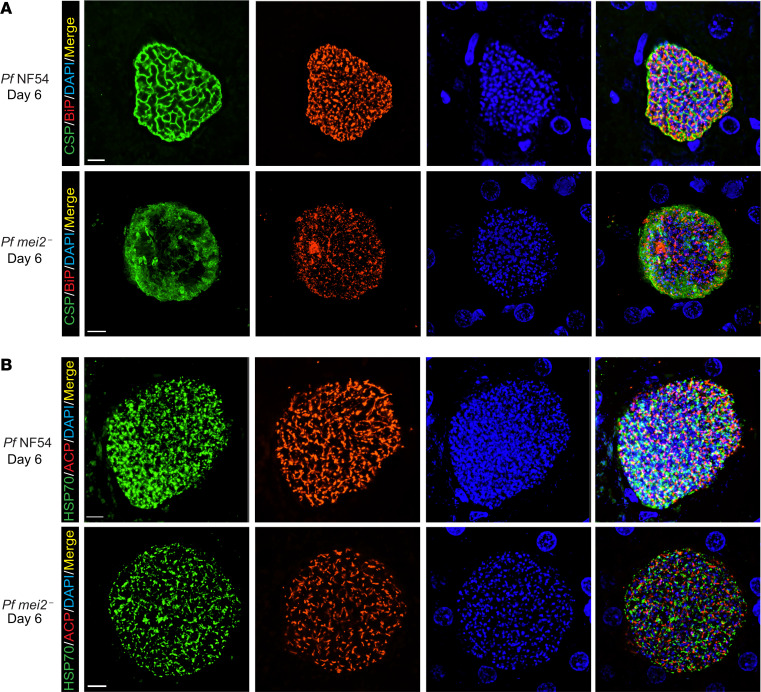
Figure 5. P. falciparum mei2– late liver stages display defects in cytomere formation.
Liver tissue sections from FRG NOD huHep mice infected with P. falciparum NF54 or P. falciparum mei2– sporozoites were prepared on day 6 of liver stage development and used for IFA. (A) IFA using an antibody against CSP (green), which delineates the PPM of P. falciparum NF54 day 6 liver stages (upper panel), shows membrane invaginations typical of cytomere formation. CSP-positive PPM invaginations are lacking in P. falciparum mei2– liver stages (lower panel). The ER was delineated using an antibody against BiP (red) and was aberrant in P. falciparum mei2–. DNA was localized with DAPI (blue). (B) IFAs were carried out to compare development of parasite organelles, including the mitochondria (mHSP70, green) and apicoplast (ACP, red), between P. falciparum NF54 (upper panel) and P. falciparum mei2– (lower panel) on day 6. The mitochondria and apicoplast development appear normal in P. falciparum mei2– day 6 liver stages. Scale bar: 7 μm.

