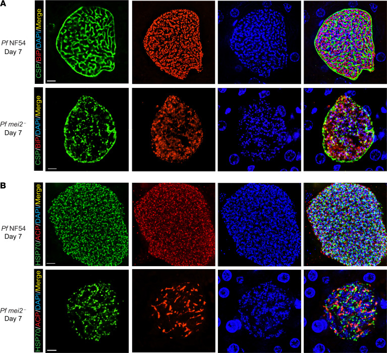
Figure 6. P.falciparum mei2– day 7 liver stages display severe differentiation defects and cannot form exoerythrocytic merozoites.
Liver tissue sections from FRG NOD huHep mice infected with P. falciparum NF54 or P. falciparum mei2– sporozoites were prepared on day 7 of liver stage development and used for IFA. (A) IFAs for CSP (green), BiP (red), and DNA (blue) in P. falciparum NF54 day 7 liver stages show further maturation and formation of distinct PPM invaginations preceding the development of exoerythrocytic merozoites (upper panel) while CSP localization for P. falciparum mei2– is aberrant (lower panel). In P. falciparum NF54 there is pronounced segregation of the ER in preparation for exoerythrocytic merozoite formation, but this does not occur in P. falciparum mei2–. (B) IFAs comparing development of the organelles mitochondria (mHSP70, green) and apicoplast (ACP, red) between NF54 (upper panel) and P. falciparum mei2– (lower panel) on day 7. The mitochondria and apicoplast have undergone segregation inside exoerythrocytic merozoites in P. falciparum NF54, while in P. falciparum mei2– they remain branched and do not segregate. Scale bar: 7 μm.

