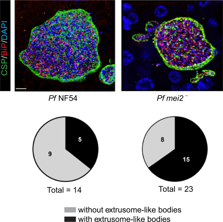
Figure 9. Extrusome-like bodies were observed in P. falciparum mei2– late liver stage schizonts.
Liver tissue sections from FRG NOD huHep mice infected with P. falciparum NF54 or P. falciparum mei2– sporozoites were prepared on day 7 of liver stage development and used for IFA using antibodies against the PPM (CSP, green), ER (BiP, red), and DNA (DAPI). The pie chart shows the distribution of late liver stage schizonts with and without presence of extrusome-like bodies. Scale bar: 7 μm.

