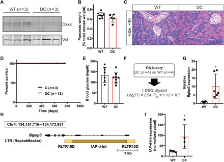
Fig. 1. Daxx loss is well tolerated in the developing pancreas.

(A) Western blot analysis of Daxx protein expression in the pancreas of wild-type (WT) or Daxxfl/flPdx1-CreTg (DC) mice with vinculin (Vcl) as a control. Each lane is one independent pancreas. (B) Pancreas weight presented as a percentage of total mouse weight (mean ± SD). (C) Representative hematoxylin and eosin (H&E)–stained sections of WT and DC mouse pancreases. Image magnification is ×40; scale bars, 100 μm. (D) Kaplan-Meier survival analysis, compared with Pdx1-CreTg (C) controls. (E) Nonfasting blood glucose measurements of 12-month-old mice (mean ± SD). (F) Schematic representation of basal RNA-seq analysis. DEG, differentially expressed gene. (G) Quantitative reverse transcription PCR validation of Bglap3 expression, shown as mean ± SD. *P < 0.05, Student’s t test. (H) Schematic representation of the murine Bglap3 locus, with intragenic ERV, IAP-d-int, flanked by RLTR10D LTRs. (I) Normalized read counts for IAP-d-int from RNA-seq data (mean ± SD). *P = 0.03, Wilcoxon test.
