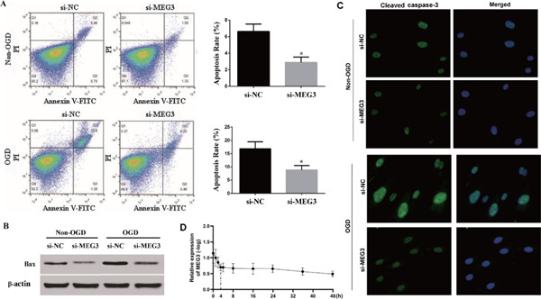
Fig. 2.

Apoptosis of human brain microvascular endothelial cells (hBMECs) in response to various disturbances (A). Apoptosis of hBMECs in response to various disturbances assessed by flow cytometry. (B). Images of western blot showed that oxygen– glucose deprivation (OGD) treatment up-regulated the expression level of Bax, while it was reversed by si- maternally expressed gene 3 (MEG3). (C). Images of immunofluorescence showed that OGD treatment up-regulated the expression level of cleaved caspase-3, while it was reversed by si-MEG3. (D).The expression pattern of MEG3 after OGD treatment.
