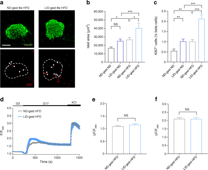
Fig. 5.
Metabolic stress-induced changes in beta cell proliferation and calcium fluxes are comparable between offspring from hypothyroid and euthyroid mothers. Adult male offspring (16–18 weeks of age) were analysed. (a) Representative confocal images of pancreatic islets in male offspring (scale bar, 100 μm, 5 μm Z-projection; red, Ki67; green, insulin). Dashed circles delineate islets and arrowheads indicate Ki67+ beta cell nuclei. (b) Quantification of islet area (n = 5–8 mice/group, one-way ANOVA). (c) Quantification of beta cell proliferation (measured as % of beta cells positive for Ki67) (n = 5–8 mice/group, one-way ANOVA). (d–f) Mean traces (d) and summary bar graphs (e, f) showing no changes in the amplitude of 17 mmol/l glucose- (e) and glucose +10 mmol/l KCl- (f) stimulated Ca2+ rises in LID-gest offspring fed HFD (n = 58–76 islets/6–7 mice/group, Mann–Whitney U test). *p < 0.05, **p < 0.01, ***p < 0.001 using the tests indicated above. Data are mean ± SEM. ND-gest ND, ND during gestation then ND; LID-gest ND, LID during gestation then ND; ND-gest HFD, ND during gestation then HFD; LID-gest HFD, LID during gestation then HFD

