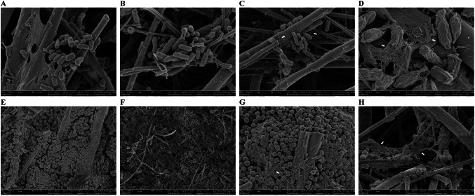
Fig. 3.
Field-emission scanning electron microscopy (FE-SEM) micrographs of Escherichia coli O157:H7 (A–D) and Staphylococcus aureus (E–H) on the surfaces of glass fiber filters contaminated as adhered cells (A and E), type I biofilms formed by immersion of inoculated glass fiber filters in TSB for 3 days at 28 °C (B and F), type II biofilms formed by cultivation of inoculated glass fiber filters on TSA for 1 days at 28 °C (C and G), or type III biofilm formed by cultivation of inoculated glass fiber filters on TSA under 100% RH for 3 days at 28 °C (D and H). Magnification: (A–C and E–G), × 20,000; (D and H), × 30,000

