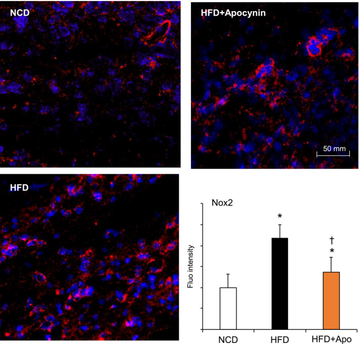
FIGURE 6.

Brain Nox2 expression detected by immunofluorescence. Nox2 was labelled by Cy3 (red) and nuclei were labeled with 4′,6‐diamidino‐2‐phenylindole4′,6‐diamidino‐2‐phenylindole (DAPI, blue) to visualized cells. Nox2 fluorescence intensities were quantified. *P < .05 for indicated values vs NCD values. †P < .05 for indicated values vs HFD values. n = 6 mice/group and at least three sections per mouse brain were used. Nox2, Nox2‐containing NADPH oxidase
