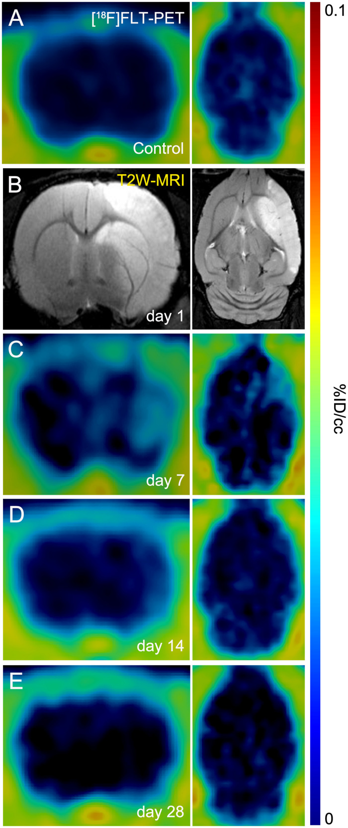
FIGURE 1.
Positron emission tomography (PET) images of [18F]FLT and Magnetic resonance imaging (MRI) [T2-weighthing (T2W)] before (control) and at days 1, 7, 14, and 28 after cerebral ischemia in rats. [18F]FLT PET (A, C–E) and MRI-T2W (B) images of axial and coronal planes at the level of the lesion are shown. Images correspond to representative animals for each time point.

