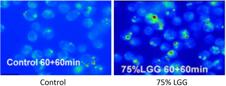
FIGURE 3.
Killing of intracellular bacteria within J774 macrophages in the presence of LGG supernatant. Viable bacteria were observed following phagocytosis using acrdine orange and crystal violet staining assay. Microscopy images show control (left, no treatment), 75% LGG (right image) and are presented as pseudo-colored fluorescence images at 60 min time points during the killing phase period. Viable bacteria are shown in green and dead bacteria are shown in red.

