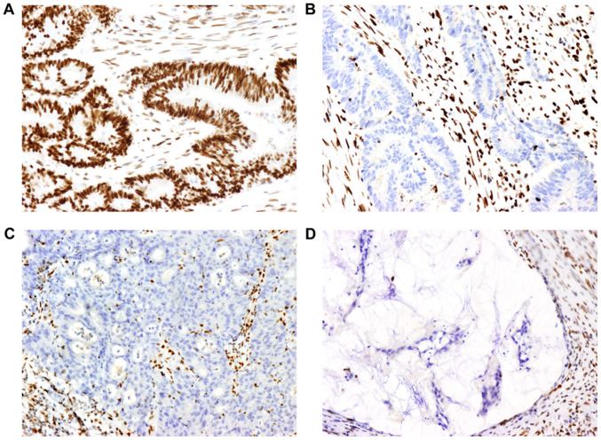
Figure 1.
Images of MMR proteins assessed by immunohistochemistry on paraffin-embedded colorectal cancer specimens. (A) Positive expression of MLH1 protein (magnification, ×200). (B, C and D) Lack of expression of (B) MSH2, (C) MSH6 and (D) PMS2 protein (magnification, ×200). Positive expression is defined as the MMR protein being expressed in the tumor cell nucleus. A negative result is defined as the absence of nuclear staining detected in tumor cells, while the surrounding stromal cells and lymphocytes present with staining and serve as an internal positive control. MMR, mismatch repair; MLH1 mutL homolog 1, DNA mismatch repair protein MLH, putative; MSH2, mutS homolog 2; MSH6, mutS homolog 6; PMS2, PMS1 homolog 2, mismatch repair system component.

