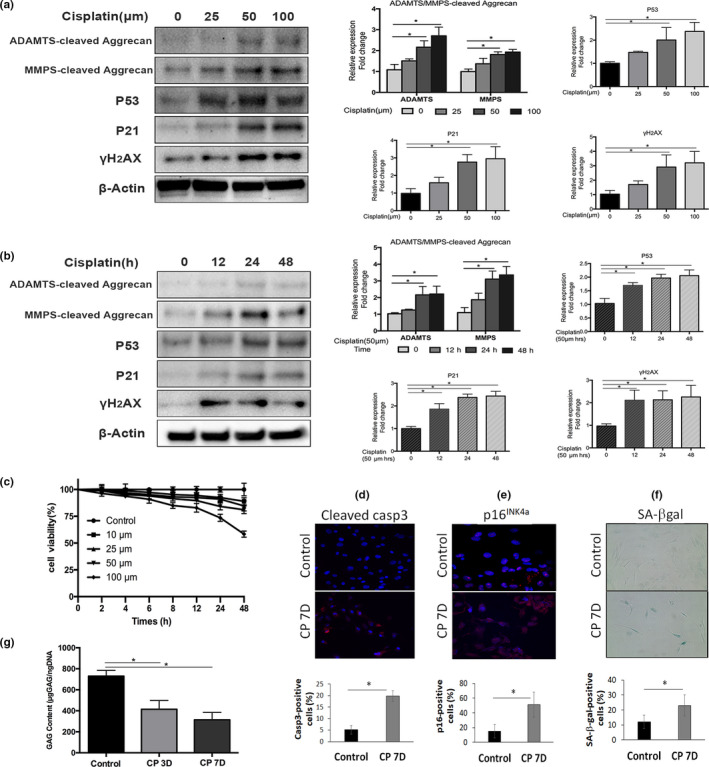
Figure 1.

Cisplatin exposure induces cellular senescence and matrix catabolism in human nucleus pulposus cells. Western blot analyses of human nucleus pulposus (hNP) cells treated with different doses (a) and durations (b) of cisplatin for the senescence markers p53, p21 and γH2AX and for the G1 containing aggrecan fragments from the matrix metalloproteinases (MMP)‐ and ADAMTS‐mediated proteolytic cleavage within the interglobular domain (IGD) of aggrecan. Aggrecan fragments shown were generated from MMP‐mediated cleavage (~55kDa) and ADAMTS‐mediated cleavage (~65kDa) of aggrecan IGD. Protein levels were normalized against β‐actin, and graph shows average values ± SD. (n = 3), *p < .05 versus. control. (c) Cell viability was determined by the CCK‐8 assay on hNP cells treated with different concentrations of cisplatin up to 48 hr. The number of viable cells decreased in a dose‐ and time‐dependent manner. (d) Apoptosis was assayed by cleaved caspase 3 immunofluorescence. (e) Cellular senescence was assayed by p16INK4a immunofluorescence. (f) SA‐β‐gal activity staining (100x magnification) of hNP cell culture treated with cisplatin for 24 hr and then returned to normal media for 7 days. Graph shows the percentage of SA‐β‐gal‐positive cells. (g) Dimethylmethylene Blue assay for total glycosaminoglycan of hNP cells treated with cisplatin for 24 hr and then returned to normal media for 3 days (CP3D) and 7 days (CP7D)
