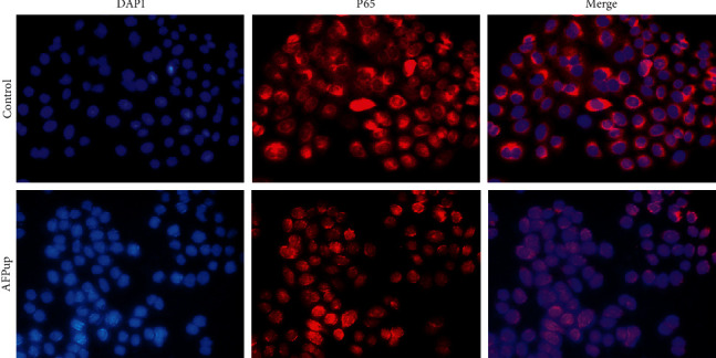
Figure 6.

Immunofluorescence analysis revealed that the majority of the P65 protein (red) was in the cytoplasm of cells from the subcutaneous hepatocellular carcinoma (HCC) xenografts transfected with negative control lentivirus and inoculated in BALB/c nude male mice. The P65 protein was translocated in the nucleus (blue) in the cells of the HCC xenografts transfected with recombinant AFP lentivirus.
