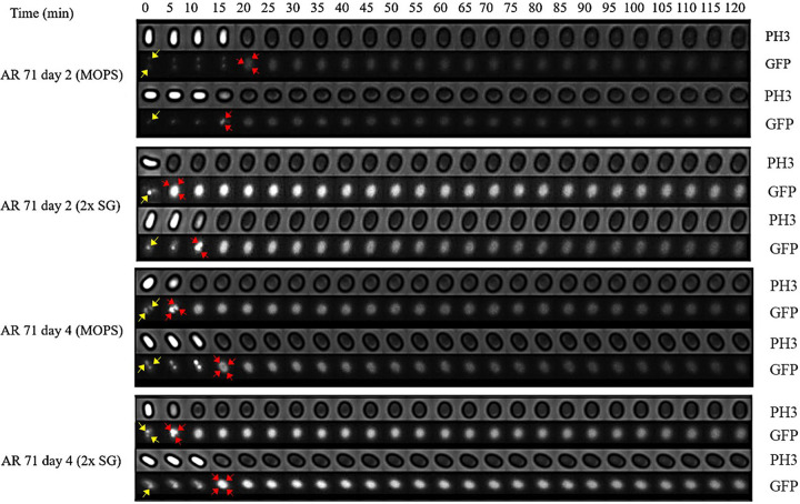
FIG 2.
Phase-contrast (PH3) and fluorescence (GFP) images of germinating B. subtilis AR71 (MalS-GFP) young and mature spores prepared on solid and liquid media. Images of individual spores, showing GFP foci (yellow arrows), were captured every 5 min for 2 h to observe the phase-contrast transition (bright to dark) and changes in MalS-GFP fluorescence intensity. The diffuse GFP foci are shown by red arrows. As representatives of the data set, only two spores per sporulation condition are shown.

