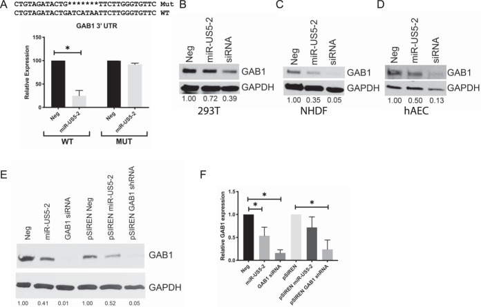
FIG 1.
GAB1 is a target of HCMV miR-US5-2. (A) The GAB1 3′ UTR or the same 3′ UTR lacking the miR-US5-2 target site was cloned into a Dual-Luciferase reporter vector and transfected into HEK293T cells along with negative-control (Neg) miRNA or miR-US5-2 mimic. 24 h later, cells were lysed, and luciferase expression was measured. Experiments were performed in triplicate. MUT, mutant. (B to D) HEK293T cells (B), normal human dermal fibroblasts (NHDF) (C), and human aortic endothelial cells (hAEC) (D) were transfected with negative-control miRNA, miR-US5-2 mimic, or a GAB1 siRNA. 48 h later, cells were lysed and subjected to immunoblotting for GAB1 and GAPDH. GAB1 band intensity was calculated using ImageJ software and compared to GAPDH band intensity. The ratio of GAB1 band intensity to GAPDH band intensity was set to 1 for the Neg sample, and each subsequent sample ratio is presented as a multiplier of the value corresponding to the Neg time point. (E) HEK293T cells were transfected with negative-control miRNA, miR-US5-2 or a GAB1 siRNA, or expression vectors expressing the miR-US5-2 hairpin or an shRNA targeting GAB1. 48 h later, cells were lysed and immunoblotted as described for panels B to D. (F) HEK293T cells were transfected as described for panel E, and RNA was harvested 48 h later. GAB1 mRNA expression levels were determined using qRT-PCR and normalized to 18S expression levels. Experiments were performed in triplicate. Data are presented as standard errors of the means. *, P < 0.05 (as determined by two-tailed two-sample t test).

