FIG 5.
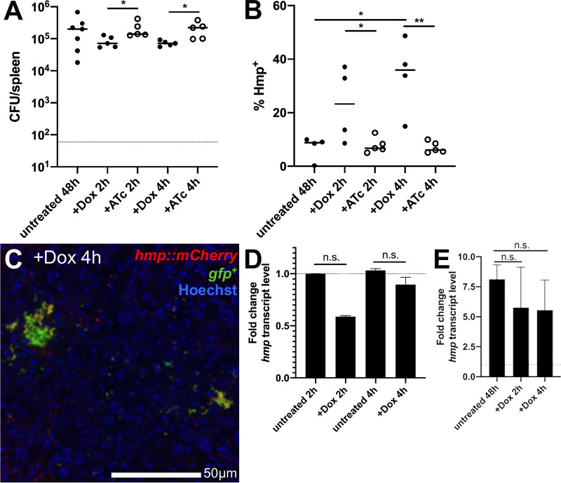
Hmp+ cells preferentially survive doxycycline treatment. C57BL/6 mice were inoculated i.v. with 103 GFP+ hmp::mCherry Y. pseudotuberculosis. Spleens were harvested from mice at 48 h p.i. (untreated). Additional mice were treated at 48 h p.i with either Dox or ATc, and spleens were harvested at the indicated time points posttreatment to (A) quantify CFU and (B) quantify the percentage of Hmp+ cells by flow cytometry. Dots indicate individual mice. Experiments performed in triplicate. (C) Representative image. (D) WT bacteria were incubated at 37°C for 2 or 4 h, with or without 0.1 μg/ml Dox. RNA was isolated, and qRT-PCR was used to detect hmp transcript levels relative to 16S rRNA levels. Fold change was calculated relative to untreated samples; median and range are shown for biological triplicates. (E) C57BL/6 mice were infected with WT Y. pseudotuberculosis, and spleens were harvested from mice at 48 h p.i. (untreated). Additional mice were treated at 48 h p.i with Dox, spleens were harvested at 2 and 4 posttreatment to isolate RNA, and qRT-PCR was performed to detect hmp transcript levels relative to 16S rRNA levels. Fold change calculated relative to hmp levels in the inoculum. n = 3 for untreated groups, n = 4 for treated groups; median and range are shown. Statistics: Kruskal-Wallis one-way ANOVA with uncorrected Dunn’s test in panels A, B, and E, and Wilcoxon matched pairs in panel D. *, P < 0.05; **, P < 0.01; n.s., not significant.

