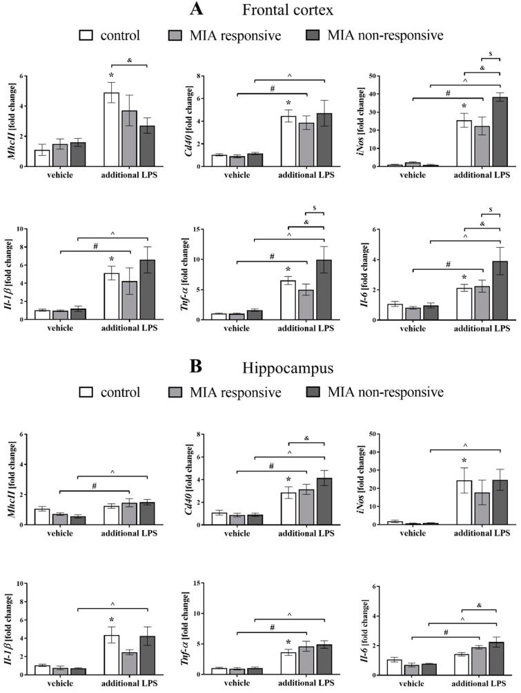
Figure 9.
The effect of MIA and the additional acute challenge with LPS on the gene expression of the pro-inflammatory microglial markers: MhcII, Cd40, iNos, Il-1β, Tnf-α and Il-6 in the frontal cortices (A) and hippocampi (B) of male Sprague-Dawley offspring at PND120. The mRNA levels were measured using qRT-PCR with n = up to 10 in each group. The results are presented as the average fold change ± SEM. * p < 0.05 vs. control + vehicle, # p < 0.05 vs. MIA responsive + vehicle, ^ p < 0.05 vs. MIA non-responsive + vehicle, $ p < 0.05 vs. MIA responsive + LPS, & p < 0.05 vs. control + LPS.

