Figure 1.
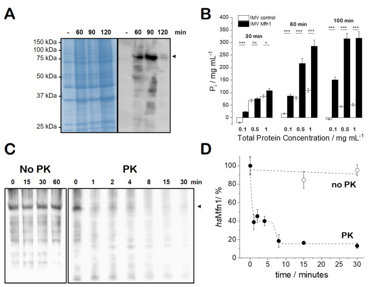
Characterisation of histidine-tagged full length human Mfn1 protein produced inE. coliSF100(DE3) strain. (A) 12% SDS-PAGE (left) and Western blot (right) with E. coli SF100(DE3)+/pTL2 cells before (-) or after (60, 90 120 min) after the addition of 1 mM IPTG. The arrowhead indicates the position of hsMfn1 (∼ 82 kDa); (B) GTPase activity of Mfn1-IMVs supplemented with 10 mM GTP and incubated as indicated at 37 °C before developing with malachite green GTPase hydrolysis assay of E. coli IMVs in the presence of 10 mM of GTP. IMVs with a total protein concentration of 0.1, 0.5 and 1 mg/mL in the absence (white bars; control) and presence of hsMfn1 (black bars) were incubated for 30, 60 and 100 min at 37 °C. Student’s t-test was performed to measure the significance of statistical difference between the different groups. and were considered statistically significant. (C) Protease protection assay of Mfn1-IMVs at a total protein concentration of 1 mg/mL in the absence (No-PK) or presence of 0.1 mg/mL of PK. IMVs No-PK were incubated at 37 °C and IMVs PK were incubated at 4 °C to be able to follow the breakdown. (D) Time evolution of protein breakdown as quantified from the protein band intensity during PK treatment.

