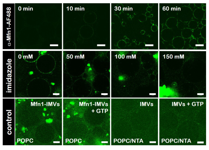
Figure 3.
Specific binding ofhsMfn1 protein to giant unilamellar vesicles (GUVs). Confocal fluorescence microscopy images showing the time series of the binding (top row) and release (middle row) of -Mfn1-488 antibody to hsMfn1 immobilised on the surface of POPC/DOGS-NTA GUVs. Prior to the incubation with POPC/DOGS-NTA GUVs, hsMfn1 itself was incubated with -Mfn1-488 (green signal). To release the bound hsMfn1 from the POPC/DOGS-NTA GUVs, increasing amounts of imidazole were added to the sample. Control assays (bottom row), where POPC GUVs (in the absence of DOGS-NTA) were incubated with Mfn1-IMVs (bottom row, Mfn1-IMVs) or POPC/NTA GUVs were incubated with solubilised E. coli IMVs (bottom row, IMVs) showed no labelling of the GUV contour upon incubation with -Mfn1-488. To visualise the presence of GUVs in the fluorescence images of the control experiments, the raw data were not corrected for signal-to-noise ratios. The addition of GTP to these samples did not promote adhesion of vesicles. Scale bars are 10 μm. Each control was repeated independently at least three times.

