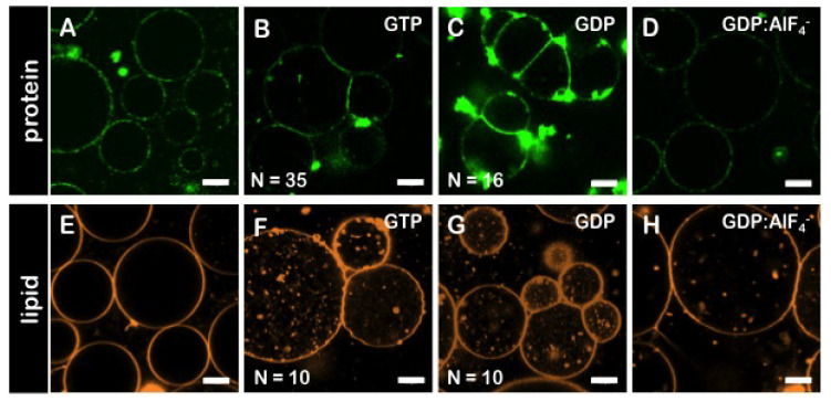
Figure 4.
Membrane adhesion of GUVs promoted byhsMfn1. Confocal fluorescence micrographs of GUVs incubated with solubilised E. coli inner membranes carrying hsMfn1 and the -Mfn1-488 antibody (top row, protein channel). (A) in the absence of nucleotide, (B) in the presence of GTP, (C) GDP and (D) GDP:AlF. The GDP:AlF complex did not promote vesicle adhesion. Similar results were obtained with GUVs labeled with RhoPE in the absence of -Mfn1-488 and incubated with solubilised Mfn1-IMVs (bottom row, lipid channel). (E) in the absence of nucleotide, (F) in the presence of GTP, (G) GDP and (H) GDP:AlF. The lipid composition of GUVs was POPC/DOGS-NTA 80/20 mol% ratio. Scale bars are 10 μm. Each condition was repeated independently at least 3 times.

