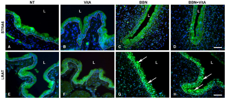
Figure 7.
Immunofluorescence of proteins stimulated by retinoic acid 6 (STRA6) and lecithin retinol acyltransferase (LRAT). (A–D) STRA6 labelling (green) is strong in the cytoplasm of urothelial cells of all groups. Scale bar = 50 μm. LRAT labelling (green) is present in the cytoplasm of the normal urothelial in (E) NT and (F) VitA groups. In (G) BBN and (H) BBN + VitA groups, LRAT labelling is strongest in the nuclei of urothelial cells (arrows). White line depicts the location of basal lamina, L, lumen. Scale bar = 50 μm.

