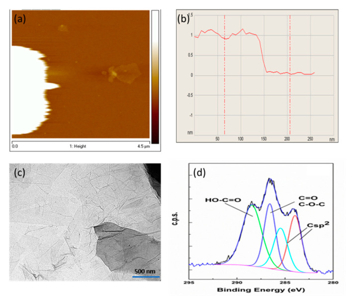
Figure 2.
Exfoliated graphene oxide (GO) monolayer atomic force microscopy (AFM) image by non-contact mode (a) and vertical height of the sheet (b); (c) TEM image (scale bar 500 nm); (d) Experimental, high-resolution XPS C1s peak of the GO employed in the present study and its best deconvolution fit to individual components.

