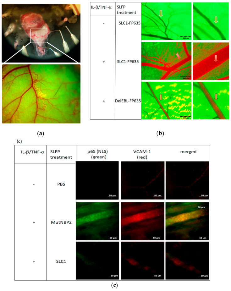
Figure 7.
In vivo imaging to visualize SLC1 binding and inhibition of NF-kappaB activation. (a) Skin flap preparation of GFP expressing transgenic mice under anaesthesia. In vivo images of the vasculature in a selected area of the tissue are depicted at different magnifications ranging from ×1.6 to ×16 and a field of view ranging from 6.9 to 0.69 mm. (b) Visualization of vessels in a non-activated state (upper row) exhibits the GFP-background fluorescence but no staining by the systemically administered red-fluorescent SLC1-FP635. Mice receiving a local cytokine-challenge (lL-1β/TNF-α) showed an intense orange staining (arrow) of the vascular endothelium (middle row) after systemic SLC1-FP635 treatment, whereas in DelEBL treated mice only a green fluorescence of the endothelial border is present (lower row). (c) Whole-mount immunofluorescence stainings of cytokine-activated mouse skin. Mice were treated intraperitoneally (i.p.) with PBS, MutNBP2 or SLC1. Whole mount was stained with an anti-NF-kappaB-(NLS) p65 antibody (green) detecting nuclear p65 and an anti-VCAM-1 antibody (red). NLS-p65 exposure and VCAM-1 expression is not detectable in unactivated (-IL-1β/TNF-α ) PBS-treated mice (upper row). NLS-p65 and VCAM-1 is detectable in activated (+ IL-1β/+TNF-α ) mice treated with the control MutNBP2 (middle row). SLC1 treatment suppresses NF-kappa activation in activated mice. Here the NLS-p65 region is not detectable accompanied by reduced endothelial VCAM-1 expression (lower row) (Figures from [23], kindly provided by PNAS).

