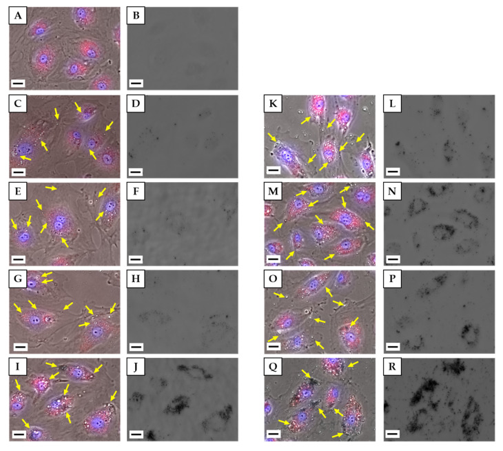
Figure 4.
Live HLEC images after incubation with H33342 for nuclear staining and CytoPainter LysosomalStaining Kit for lysosomes. (A,B) Control, (C,D,K,L) unCNH, (E,F,M,N) oxCNH, (G,H,O,P) CB, (I,J,Q,R) MWCNT. (A,C,E,G,I,K,M,O,Q) Image obtained by merging the phase contrast image and the fluorescence images. (B,D,F,H,J,L,N,P,R) Bright field image. (C–J) 10 µg/mL. (K–R) 100 µg/mL. Scale bars correspond to 20 μm. Yellow arrows indicate CNMs. HLEC, human lymphatic endothelial cell; H33342, bisbenzimide H33342 fluorochrome trihydrochloride; unCNH, untreated carbon nanohorn; oxCNH, oxidized carbon nanohorn; CB, carbon black; MWCNT, multi-walled carbon nanotube; CNMs, carbon nanomaterials.

