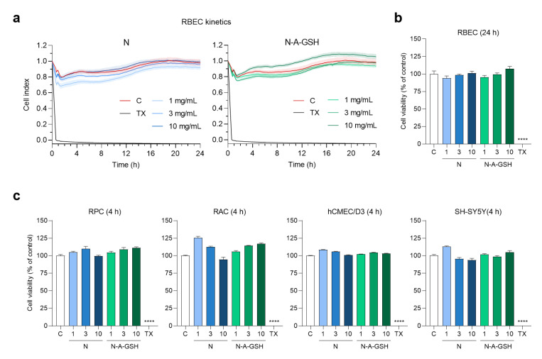
Figure 3.
Effect of non-targeted (N) and alanine-glutathione dual-targeted (N-A-GSH) niosomes at concentrations of 1, 3 or 10 mg/mL on the viability of NVU cells monitored by real-time impedance measurement. (a) Kinetics of rat brain endothelial cell responses to niosome treatments for 24 h. Values presented are means ± SEM and are given as cell index. (b) Rat brain endothelial cell viability after 24 h. (c) Cell viability of pericytes (RPC), astrocytes (RAC), hCMEC/D3 endothelial cells and SH-SY5Y neurons after 4 h. Values presented are means ± SEM and are given as a percentage of control. Statistical analysis: one-way ANOVA followed by Dunnett’s posttest; **** p < 0.0001 compared to the control group; n = 6–8. C: culture medium-treated control group; TX: Triton X-100 reagent, indicating maximum cellular toxicity.

