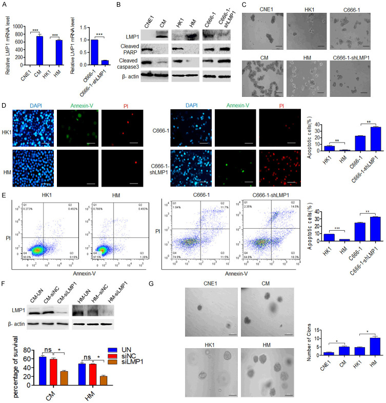
Figure 1.
LMP1 promotes anoikis resistance. (A) qPCR was performed to determine the expression of LMP1 in NPC cells (CNE1, CM, HK1, HM, C666-1, and C666-1-shLMP1 cells). (B) Western blot was used to analyze LMP1, cleaved PARP and caspase3 levels in NPC cells after suspension culture for 48 h. (C) NPC cells were grown under anchorage-free conditions (in ultralow-attachment cell culture plates) for 24 h. Scale bar: 200 μm. (D) Fluorescence microscopy and (E) flow cytometric analysis were performed to observe cell apoptosis after culture under suspension conditions for 24 h. Scale bar: 50 μm. (F) After transfection with siRNA-NC or siRNA-LMP1, the viability of nonadherent cells was assessed in a trypan blue rejection experiment. Scale bar: 100 μm. (G) The colony numbers of NPC cells in soft agar (2 weeks) are shown. Scale bar: 100 μm. CM (CNE1-LMP1), HM (HK1-LMP1). UN: untreated, NC: negative control. Columns: mean of three replicates; statistical significance was calculated using a t-test (ns: no significance, *P<0.05, **P<0.01, and ***P<0.001).

