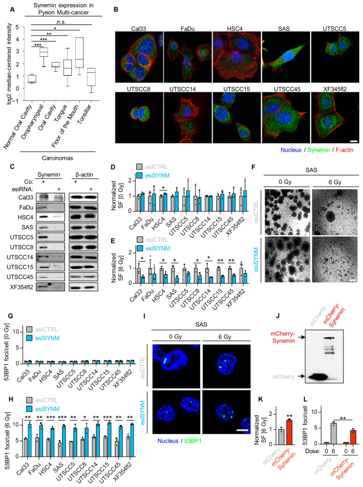
Figure 2.
Synemin essentially controls radiosensitivity and DSB repair. (A) Analysis of synemin mRNA expression in head and neck carcinomas in comparison to corresponding normal tissue using Oncomine database; (B) Immunofluorescence staining of synemin distribution (green) in a panel of HNSCC cell lines. Cells were counterstained with Phalloidin (F-actin, red) and DAPI (nucleus, blue) (bar, 20 µm); (C) Immunoblots with knockdown efficiencies in a panel of HNSCC cell lines; (D) Normalized plating efficiency of a panel of HNSCC cell lines upon synemin inhibition (n ≥ 3); (E) Colony formation ability of 6-Gy X-ray irradiated 3D lrECM HNSCC cell cultures after esiRNA-mediated synemin depletion; (F) Representative phase contrast images of 3D lrECM SAS cell cultures (bar, 500 µm); (G) Spontaneous foci per cell in a panel of HNSCC cell lines upon synemin inhibition (n = 3); (H) Effect of synemin silencing on residual 53BP1 foci (24 h after irradiation) in a panel of 6-Gy irradiated 3D lrECM HNSCC cell lines; (I) Representative immunofluorescence images of residual 53BP1 foci (bar, 10 µm); (J) Immunoblot of mCherry–Synemin and mCherry empty vector expression; (K) Colony formation ability of SAS mCherry–Synemin transfectants, relative to SAS mCherry controls (6-Gy X-rays); (L) Residual 53BP1 foci (24 h after irradiation) in SAS mCherry–Synemin transfectants exposed to 6-Gy X-rays. Data are presented as mean ± SD (n = 3; two-sided t-test; * p < 0.05, ** p < 0.01, *** p < 0.001).

