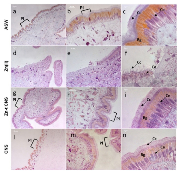
Figure 8.
Light microscopy of mussels’ mantle edges stained with haematoxylin and eosin (H&E) of specimens exposed to the following experimental groups: ASW (a–c, control); Zn(II) (d–f, 10 mg L−1 of ZnCl2 in ASW); Zn-t CNS (g–i, ZnCl2 10 mg L−1 contaminated ASW treated with CNS); CNS (l–n, ASW treated with only CNS). Cc: Ciliated columnar cells; Ce: columnar epithelium; Bg: brown intracellular granules; Pl: plicae. Images obtained with a Zeiss Axiophot epifluorescent microscope with AxioCamMRc5.

