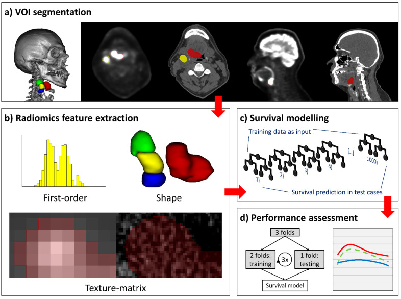
Figure 4.
Segmentation, radiomics feature extraction, and survival modelling pipeline. (a) Positron emission tomography (PET)-guided manual segmentation of the primary tumor and individual metastatic cervical lymph nodes on PET and computed tomography (CT) axial slices, sagittal images, and a 3D-renderered image are provided for spatial awareness. (b) Extraction of first-order, shape, and texture matrix features yielded 1037 radiomics features per imaging modality and per lesion. (c) Random forest machine-learning models with 1000 decision trees were applied for survival prediction and risk-stratification. (d) Model performance was assessed in threefold cross-validation (left), wherein all subjects were assigned to three folds by stratified random split; the models were trained on two folds, and one fold was used for model validation. Model performance was visualized in performance curves (right).

