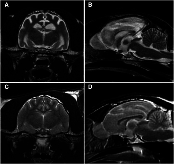
Figure 1.
Axial (A) and sagittal (B) T2-weighted magnetic resonance images of the affected cat’s brain showing loss of gray/white matter distinction in the cerebral cortex and diffuse cerebral cortical atrophy (B). Comparable axial (C) and sagittal (D) images of the brain of a healthy young adult cat.

