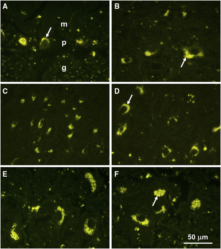
Figure 2.
Fluorescence micrographs of sections of the cerebellar cortex (A), a deep cerebellar nucleus (B), the parietal cerebral cortex (C and D), and brainstem nuclei (E and F). In the cerebellar cortex, the largest accumulations of storage bodies were present in the Purkinje cell layer (p), with scattered storage body aggregates in the granule cell (g) and molecular (m) layers.

