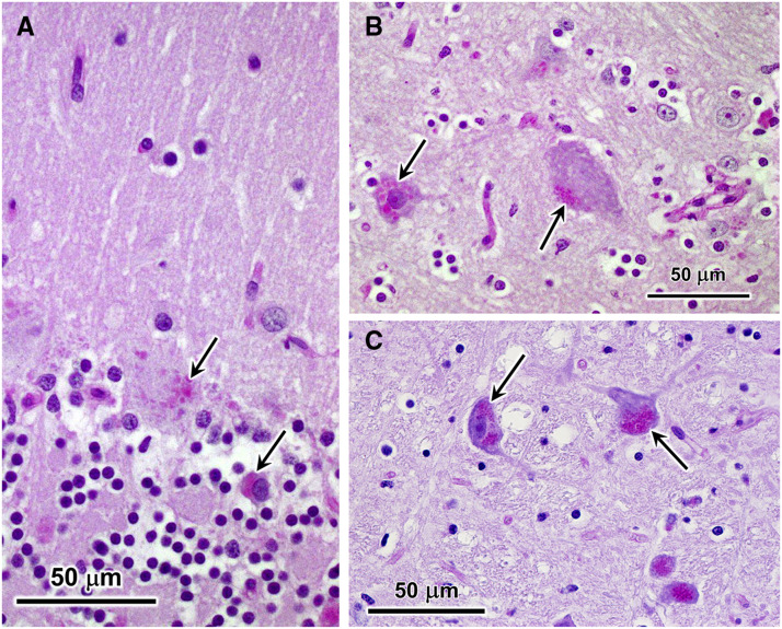
Figure 3.
Light micrographs of sections of cerebellar cortex (A), cerebral cortex (B), and brainstem (C) from the affected cat. Sections from the cerebellar cortex and cerebral cortex were stained with periodic acid-Schiff (PAS) reagent, and the section from the brainstem was stained with a combination of PAS and Luxol fast blue (LFB). Purkinje cells (top arrow in A) and occasional cells in the molecular layer (lower arrow in A) contained aggregates of PAS-stained inclusions. Similar inclusions were present in cerebral cortical neurons (arrows in B). In brainstem neurons, similar inclusions appeared purple when stained with a combination of PAS and LFB (arrows in C).

