Figure 2.
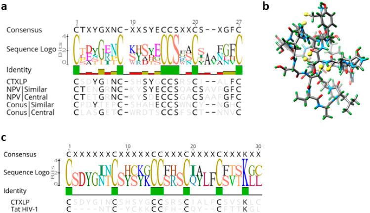
ERVK CTXLP is conserved structurally with Conus and viral conotoxins, as well as Human Immunodeficiency Virus (HIV) Tat. (a) ERVK CTXLP cysteine-rich motif has a strong similarity to both nuclear polyhedrosis virus (NPV, 46.2%) and Conus (45.8%) conotoxin proteins. Cysteine bridges conserved in knottin proteins are indicated with black bars. (b) A modeled 3D structure of the CTXLP domain was predicted using the Knotter1D3D software. Note the interactions of the yellow cysteine residues, as they form disulfide bonds. (c) Alignment and sequence logo of the cysteine-rich motif in ERVK CTXLP peptide and cysteine-rich domain of HIV-1 Tat protein. Conservation of six of the seven CTXLP cysteine residues is found in HIV Tat, as well as a C-terminal lysine residue.

