. 2020 May 19;11(24):6134–6148. doi: 10.1039/d0sc01935f

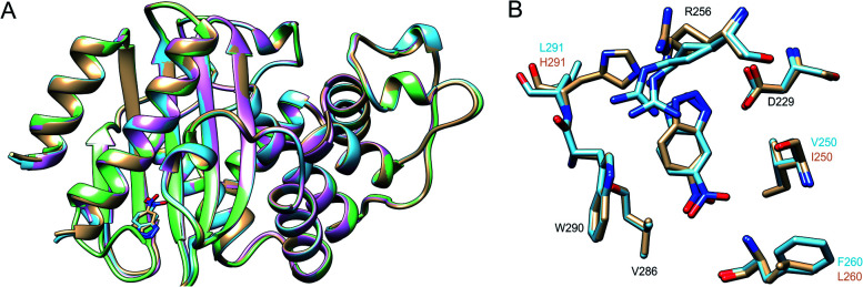
Fig. 5. (A) Superposition of the 3D crystal structures of the background GNCA4-WT (tan, PDB ID: 5FQK 35) and the three FuncLib variants whose structure we have determined in this work, specifically the GNCA4-2 (light blue, PDB ID: ; 6TY6), GNCA4-12 (pink, PDB ID: ; 6TXD) and GNCA4-19 (green, PDB ID: ; 6TWW) variants. Highlighted here is also the position of the transition state analogue in the GNCA4-WT and GNCA4-2 variants. (B) A close-up of the de novo active site in these enzymes, superimposing the active sites of the background enzyme (tan) and the GNCA4-2 variant predicted from FuncLib (light blue, Table 3), with a transition state analogue bound in the active site. Note that we have changed the orientation of the active site compared to panel (A), to better highlight the changes in key active site side chains.

Fig. 5