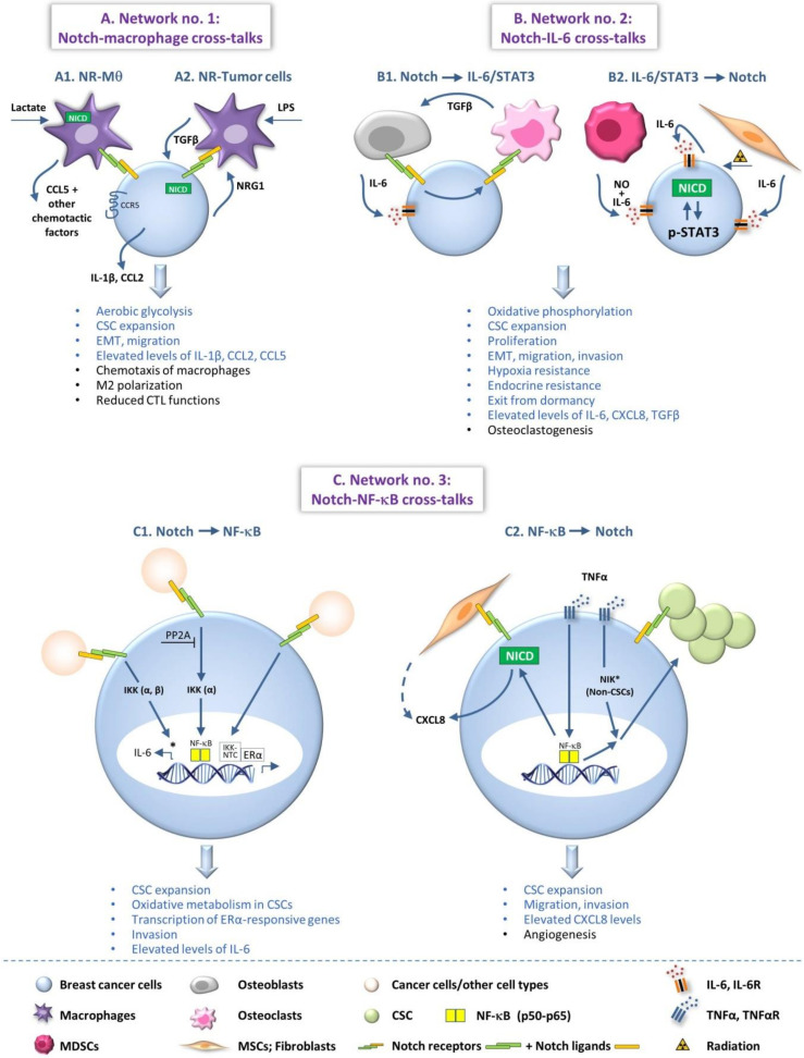
Figure 1.
Notch-inflammation networks that promote breast cancer progression. The Figure demonstrates three major networks that are established between members of the Notch pathway and inflammatory elements in BC. For the sake of simplicity, not all processes and not all mechanistic details that were described in the different studies are demonstrated in the Figure. When several processes that were identified in different publications are illustrated in the same cell/setting, there is no intention to imply that they take place simultaneously. In the text, the readers are referred to the relevant parts that are demonstrated in the Figure. Three Notch-inflammation networks are demonstrated in the Figure: (A) Network no. 1—Cross-talks between the Notch pathway and macrophages, as key representatives of myeloid cells that contribute to cancer-related inflammation; (A1) Effects of tumor-induced activation of Notch signaling in macrophages; (A2) Effects of macrophage-induced activation of Notch signaling in the tumor cells. (B) Network no. 2—Cross-talks between the Notch pathway and IL-6—a major pro-inflammatory cytokine that is one of many pro-inflammatory factors prevailing in breast tumors and promoting disease progression—and its downstream pathways (STAT3); (B1) Effects of Notch activation on IL-6/STAT3 signaling; (B2) Effects of IL-6/STAT3 signaling on Notch activation in the tumor cells. (C) Network no. 3—Cross-talks between the Notch pathway and the NF-κB transcription factors, which have prominent roles in inflammatory processes and mediate pro-inflammatory/pro-metastatic effects in many malignancies, including BC; (C1) Effects of Notch signaling through activation of the NF-κB pathway, on breast tumor cells; (C2) Effects of NF-κB-mediated effects through Notch activation on breast cancer cells. *, aspects concerning the activation of non-classical NF-κB pathways. In the texts describing the pro-tumor consequences of the Notch-inflammation networks, the blue text signifies pro-malignancy effects potentiated in the tumor cells and the black text illustrates tumor-promoting activities at the TME. Please note that factors released by the cancer cells can also affect the characteristics of the TME. CSCs, cancer stem cells; CTLs, cytotoxic T cells; EMT, epithelial-to-mesenchymal transition; ERα, estrogen receptor α; IKK, IκB kinase; IL-6, interleukin 6; IL-6R, IL-6 receptor; LPS, lipopolysaccharide; MDSCs, myeloid-derived suppressor cells; MSCs, mesenchymal stem cells; Mθ, macrophages; NICD, Notch intracellular domain; NIK, NF-κB inducing kinase; NO, nitric oxide; NR, Notch receptors; NRG1, Neuregulin 1; NTC, Notch–CSL–MAML1 transcriptional complexes; TGFβ, transforming growth factor β; TNFα, tumor necrosis factor α; TNFαR, TNFα receptor.

