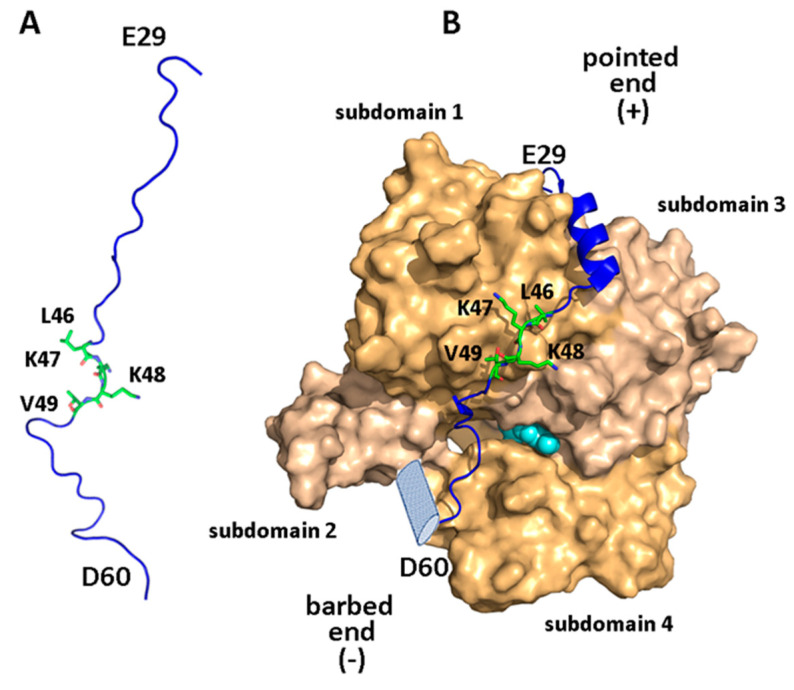
Figure 3.
Structural view of the WIP-N/actin interaction. (A) A plausible representation of residues 28–61 of WIP (blue) and sidechains of residues 46–49 in their free form, (B) bound WIP(28–61) in complex with actin (pale/dark orange, PDB ID: 2A41 [68]), showing actin subdomains. Residues of the actin-binding LKKV motif are highlighted in green. The putative C-terminal helix (absent in WIP but present in other actin-binders) is portrayed as a light-blue cylinder, and the bound nucleotide (between subdomains 3 and 4) is shown as cyan-colored spheres.

