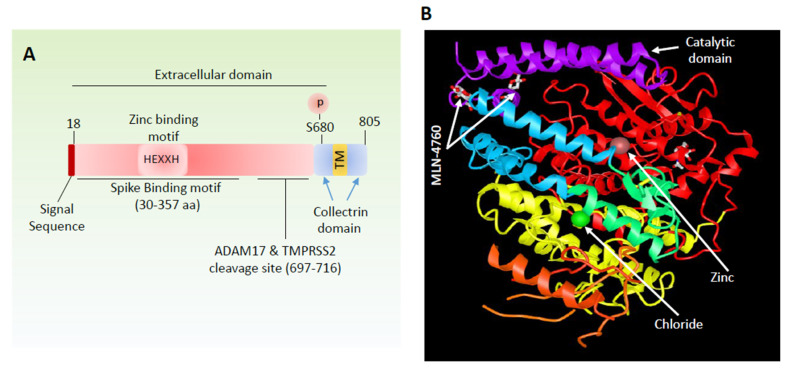
Figure 2.
Schematic and domain structure of angiotensin-converting enzyme 2 (ACE2). (A) General domain information including, ion binding, proteolytic cleavage sites and S protein binding motif are shown. (B) Crystal structure of ACE2 and location of ion bindings and catalytic domain in complex with ACE2 inhibitor, MLN-476, is shown.

