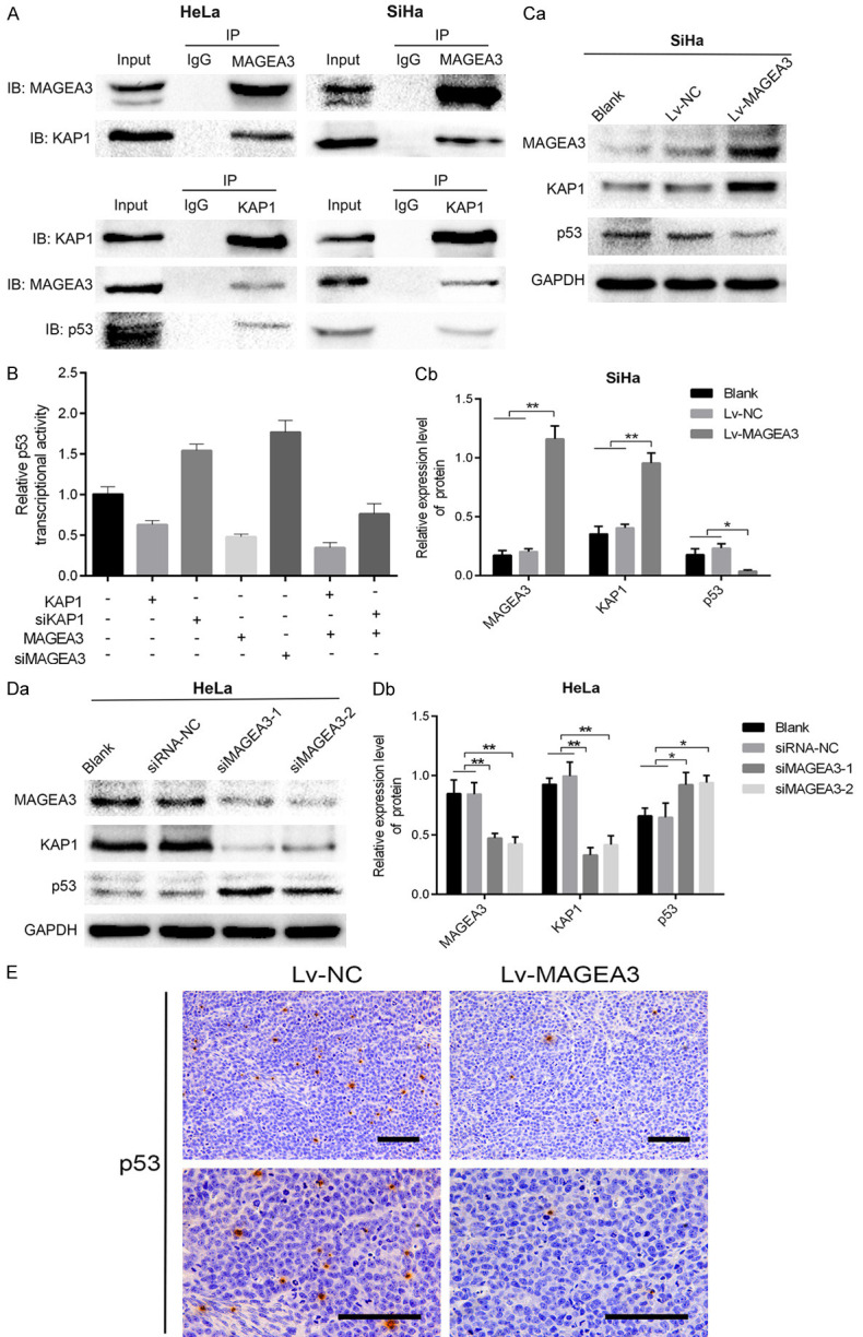
Figure 4.

MAGEA3 interacts with KAP1 and inhibits p53 transcriptional activity. A. Co-IP assays were performed in HeLa cells and SiHa cells cotransfected with Lv-MAGEA3 and pcDNA3.1-KAP1. Cell lysates were immunoprecipitated (IP) with control IgG or the indicated antibody, and the precipitated protein was detected by immunoblotting (IB) analysis with the indicated antibody. Cell extracts were used as a positive control (Input). B. Effects of MAGEA3 or KAP1 on p53 activity, as indicated by the reporter plasmid pp53-TA-luc. HeLa cells (with endogenous wild-type p53) grown on a 12-well plate were cotransfected with the pp53-TA-luc and the indicated plasmids. Luciferase was measured at 48 h posttransfection and normalized according to Renilla luciferase activities. Data (means ± SD) are represented as fold differences relative to that observed in cells without transfecting the indicated plasmids. C, D. Western blotting analysis of the p53 and KAP1 proteins following overexpression or knockdown of MAGEA3 in SiHa (Ca, Cb) and HeLa (Da, Db) cells. *P<0.05, **P<0.01 vs. control groups. E. Immunohistochemical analysis of p53 expression in the Lv-MAGEA3 group and Lv-NC group of xenograft tumors. Bar length: 100 µm.
