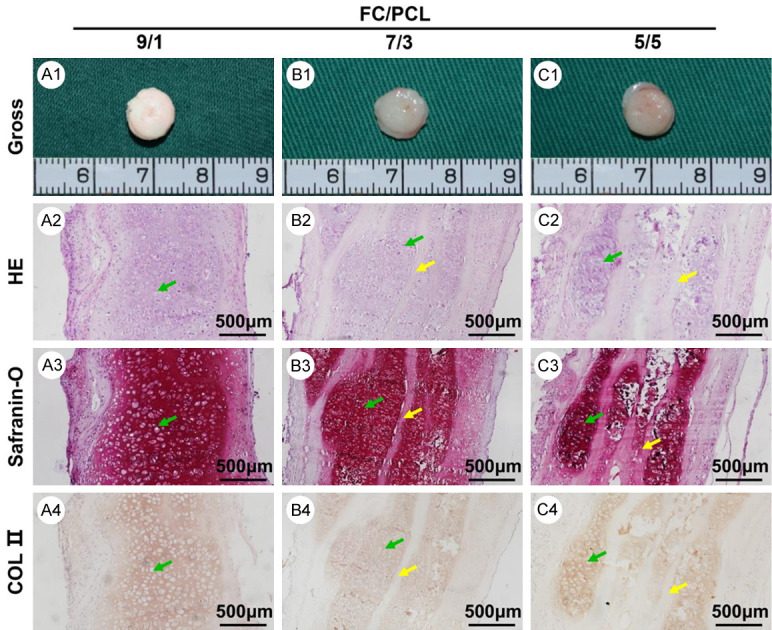
Figure 5.

In vivo neocartilage evaluation at 8 weeks. Gross view (A1-C1) of all three groups presented similar ivory-white appearances. The histological assessment, including HE (A2-C2), safranin-O (A3-C3), and collagen II (A4-C4) staining of all three samples, revealed that increases in the FC content led to a more homogeneous cartilage-specific extracellular matrix distribution and fewer residual electrospun membranes. Yellow arrows indicate undegraded electrospun membranes. Green arrows indicate neocartilage.
