Figure 4.
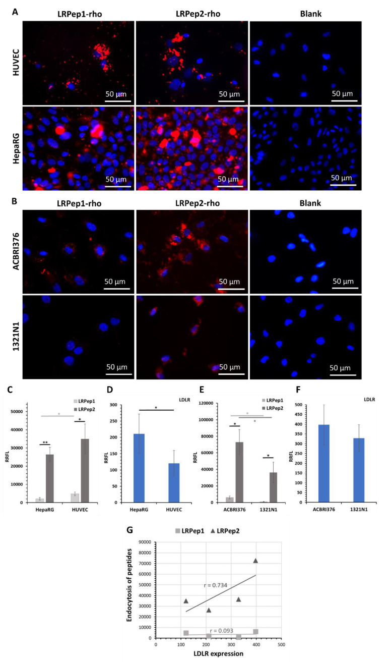
Endocytosis of the peptides LRPep1-rho and LRPep2-rho (stained in red) in (A) HUVEC and HepaRG cells and (B) ACBRI376 and 1321N1 cells. Nuclei are stained in blue with Hoechst. (C–F) The endocytosis of peptides, as well as the expression of LDLR, was semi-quantitatively evaluated by the measurement of fluorescent labeling using the ImageJ software and was normalized to the number of cells and to the background, giving the relative ratio of fluorescent labeling (RRFL); *: p < 0.05, **: p < 0.001. (G) The correlation coefficient between the expression of LDLR and the endocytosis of peptides.

